

How Cells Identify, Dismantle and Reuse Their Own Components
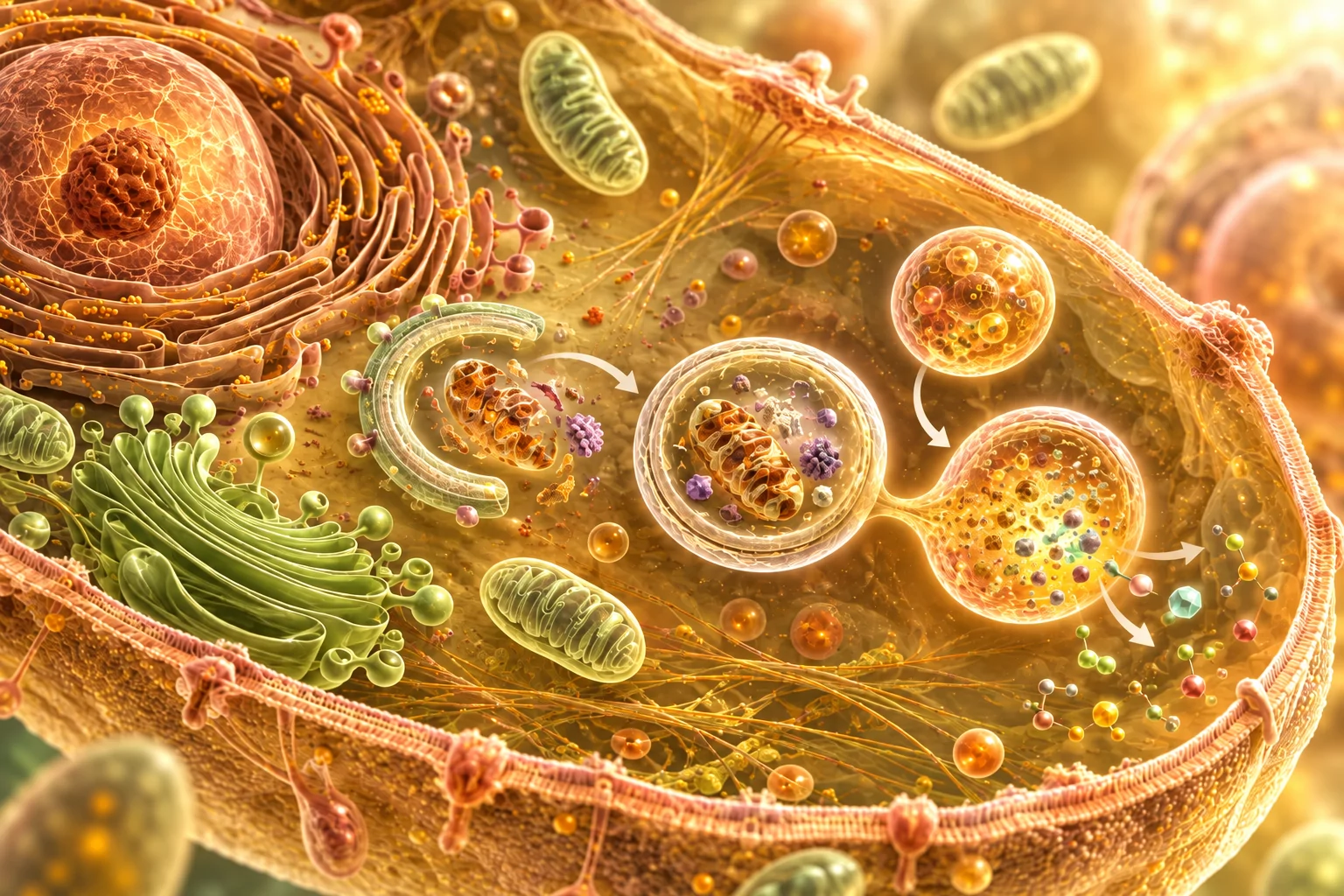
Autophagy is one of the most elegant maintenance systems in biology. The word literally means “self-eating”, yet the process is better understood as a form of controlled intracellular recycling through which cells continuously manage proteins, organelles and other structures that have aged, become damaged, lost efficiency or are no longer useful.
By isolating and dismantling part of this material, the cell can recover components that may then be reused. Autophagy is therefore part of normal cellular housekeeping and contributes both to maintenance and to adaptation when external nutrients become scarce.
From Waste to Resource
The basic sequence is highly organised.
A membrane begins to form around cellular material destined for degradation, gradually creating a double-membrane structure known as an autophagosome. Once formed, the autophagosome fuses with a lysosome, whose enzymes break down the enclosed contents.
The resulting components — including amino acids, fatty acids, sugars and other molecules — can then return to the cellular pool and be reused.
Seen in this way, autophagy is a process of recovery as much as degradation: material that has become inefficient, damaged or expendable can be converted into raw material for further cellular activity.
Why Fasting Matters
Autophagy occurs continuously, although nutrient availability strongly influences its intensity and regulation.
When nutrients and growth signals are abundant, cells tend to favour synthesis, growth and storage. As nutrient availability decreases, the balance changes: insulin and amino-acid signalling fall, mTOR activity decreases, and energy-sensing systems such as AMPK assume greater importance.
These changes create a cellular environment in which recycling and maintenance processes may become more prominent.
This is one of the principal reasons fasting has attracted so much interest in autophagy research. Calorie restriction and fasting have repeatedly been associated with increased autophagic activity in experimental models, while the extent, timing and tissue-specific response in living humans remain more difficult to measure directly.
Autophagy Is a Family of Processes
Autophagy is often presented as though the cell simply gathers random material and digests it. The reality is considerably more sophisticated.
One form, commonly called bulk autophagy, can capture portions of cytoplasmic material relatively broadly, especially during nutrient deprivation. Alongside it exists a range of selective autophagy mechanisms in which specific structures are recognised and targeted for removal.
These include:
- mitophagy, directed towards mitochondria;
- aggrephagy, involving aggregated proteins;
- ER-phagy, targeting portions of the endoplasmic reticulum;
- ribophagy, involving ribosomes;
- pexophagy, targeting peroxisomes;
- xenophagy, which can participate in the removal of invading microorganisms.
The existence of these mechanisms shows that cellular recycling can operate with a high degree of discrimination.
Mitophagy: Quality Control of the Cell’s Powerhouses
 Mitochondria deserve particular attention because they play a central role in energy production.
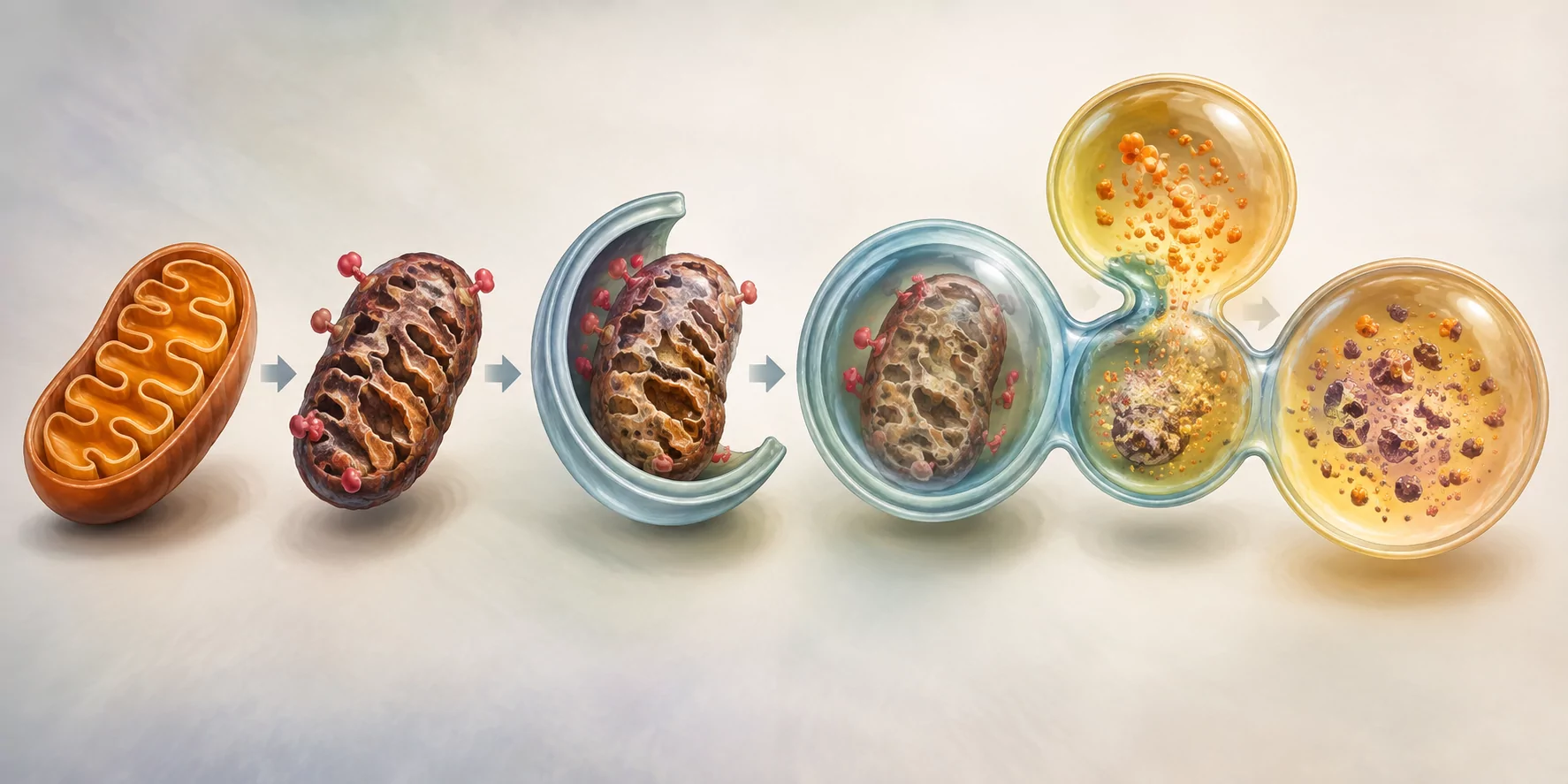
When a mitochondrion becomes damaged or inefficient, it can produce excessive reactive oxygen species and interfere with normal cellular function. Cells therefore possess mechanisms that allow dysfunctional mitochondria to be identified and removed selectively.
One of the best-studied pathways involves PINK1 and Parkin. When mitochondrial function is impaired, molecular signals can mark the affected organelle for removal, after which it is enclosed and delivered to the lysosomal system for degradation.
This process represents a form of cellular quality control in which structures that are no longer functioning adequately can be removed while healthier ones remain available.
Fasting, Repair and Cellular Priority
The relevance of autophagy becomes particularly clear during fasting, when the cell has less external material available and becomes increasingly dependent upon its internal resources.
Under these conditions, damaged or expendable components represent both a burden and a potential source of reusable material. Autophagy can therefore contribute simultaneously to maintenance, adaptation and recycling, helping the cell to preserve efficiency while recovering useful components.
This provides one modern cellular mechanism that resembles part of what older Natural Hygiene literature described more broadly as autolysis.
The two concepts should remain distinct. Hygienic autolysis referred to the internal economy of the organism as a whole, while autophagy describes specific regulated processes occurring inside living cells. Their conceptual relationship lies in the common theme of using what is already within the organism to support survival, maintenance and renewal.
Cellular “Cleaning”: A Useful Metaphor With Limits
The expression “cellular cleaning” is attractive because it immediately conveys the idea of removing damaged or unnecessary material, yet it can also oversimplify what is happening.
Autophagy participates in cellular quality control and can eliminate dysfunctional organelles, protein aggregates and other components. Its activity varies according to tissue, nutrient availability, age, stress and disease state, and its biological value depends upon regulation and context.
The useful concept is therefore one of appropriate recycling and maintenance, rather than the idea that maximum autophagy is always desirable or that fasting automatically performs a uniform “deep clean” throughout the entire organism.
When Does Autophagy Increase During a Fast?
This is one of the most common questions surrounding fasting, and also one of the areas where online explanations become unnecessarily rigid.
Autophagy is already active before fasting begins. Fasting changes the regulatory environment in which the process takes place: nutrient signalling falls, cellular energy sensing changes and pathways associated with recycling become more favourable.
The precise timing, however, varies between tissues and individuals, while direct measurement of autophagic flux in living humans remains technically difficult.
For that reason, statements claiming that autophagy “starts” at one specific hour or reaches a universal maximum after a predetermined number of fasting hours go beyond what can currently be established with precision.
The process is real; its timing is dynamic.
A Selective Intelligence
Autophagy reinforces a principle that already appears throughout fasting physiology: biological systems are capable of discrimination.
Cells can preserve some structures, repair others and selectively dismantle components that have become damaged or inefficient. Certain mitochondria may be specifically tagged for removal, while others remain untouched; protein aggregates can be recognised as targets; particular cellular structures can be recycled according to need.
This is one of the reasons the older idea of an internal intelligence remains evocative.
Modern biology describes the phenomenon through receptors, molecular recognition, signalling pathways and quality-control systems. The language is more precise, while the underlying observation remains remarkable: living systems continually distinguish between what should be preserved, what can be repaired and what may be dismantled and reused.
What We Know — and What We Are Still Learning
Autophagy is now a well-established biological process. Its role in cellular maintenance, adaptation to nutrient scarcity and selective quality control belongs firmly within modern cell biology.
What remains less clear is how far these intracellular mechanisms contribute to the broader tissue-level changes sometimes observed during prolonged fasting.
Questions remain open.
Can changes in cellular recycling contribute to the reduction of longstanding structural abnormalities? Under what conditions might this occur? Are some tissues more responsive than others? How strongly does fasting duration influence the process? And how much of what earlier practitioners described as “autolysis” can eventually be explained through autophagy and related mechanisms?
The existence of autophagy does not settle every historical claim made about fasting, although it provides a biological framework that makes some older observations considerably more interesting to investigate.
Personal Experience and Clinical Observation
My interest in autophagy is not purely theoretical. It is connected with changes that I have personally experienced during and after prolonged fasting, some of which were also documented or observed clinically.
These experiences involve different parts of the body and different kinds of change. Precisely for this reason, I find them particularly interesting in relation to the systemic nature of fasting: I could not choose where the process would act, nor could I direct it towards a particular tissue. I simply fasted, and the changes emerged where the organism appeared to have its own priorities.
Changes Visible on My Radiographs
Beginning in 2007, after a severe bilateral pleuropneumonia requiring a prolonged hospital stay, I underwent repeated chest radiographs over several years.
Because chest X-rays also include part of the vertebral column, the images repeatedly showed arthritic calcific changes along my spine.
From 2010 onwards, chest radiography also became part of my periodic maritime medical examinations required for navigation, giving me a further series of comparable images over time.
After I began undertaking prolonged fasts, something unexpected happened.
During one of these examinations, the doctor of the maritime health authority noticed that the calcifications previously visible along my spine had markedly diminished or were no longer apparent. I had not asked him to look for this change; he was the person who noticed it and pointed it out to me.
When he mentioned it, I explained that during the intervening period I had undertaken fasting.
I subsequently showed the radiographs to other doctors who had followed me previously, particularly in connection with the severe respiratory illness of 2007. They independently confirmed the same change and expressed considerable surprise, explaining that in their clinical experience spinal calcific changes were not expected to disappear spontaneously and would more commonly persist or become more evident with age.
The radiographs demonstrate that a change occurred. They cannot, by themselves, tell us which biological mechanism produced it.
That distinction is important.
The observation is documented; the mechanism remains open to investigation.
From Proposed Spinal Surgery to Full Mobility
A second experience concerns a longstanding problem at L5–S1, where I had been diagnosed with spondylolysis and spondylolisthesis.
The condition caused significant pain and functional difficulty. At Santa Corona Hospital in Pietra Ligure, Professor Valsania considered the problem serious enough to propose spinal surgery involving internal fixation with metal screws and plates.
I never underwent that operation.
After fasting, the pain disappeared, and the condition ceased to limit my everyday movement in the way it had before. Over the years that followed, I recovered a degree of physical freedom that would have seemed improbable when spinal surgery was being discussed.
Today I dance Argentine tango with great pleasure, carry out a wide variety of physical movements and activities, and am also able to move substantial weights without the spinal pain that had previously restricted me.
This functional recovery does not demonstrate that fasting anatomically reversed the spondylolysis or spondylolisthesis, nor does it identify autophagy as the process responsible.
What it does establish in my experience is a very clear before-and-after change: a spinal condition severe enough for fixation surgery to be proposed was followed, after fasting, by long-term disappearance of the symptoms that had made such intervention appear necessary.
A Third Change: The Aftermath of Pleurisy
A third experience takes me back to the severe respiratory illness of 2007.
Alongside bilateral pneumonia, I also developed pleurisy and remained in hospital for approximately twenty days.
Pleural inflammation can leave persistent structural changes after the acute illness has resolved. The inflammatory exudate is a complex biological fluid containing proteins and cells, and during healing it may be followed by fibrin deposition, adhesions, fibrosis and, in some cases, calcific changes.
In my case, the long-term consequence was very clear: any breath substantially deeper than normal produced considerable pain.
The simple mechanical act of expanding the lungs beyond ordinary quiet breathing was painful enough that I eventually required regular analgesic treatment.
After fasting, this pain disappeared.
It disappeared so completely, and remained absent for so long, that when I began reconstructing these experiences for this article I realised that I had almost forgotten how important that pain had once been in my daily life.
I no longer needed the regular pain relief that had previously been necessary, and deeper breathing ceased to produce the persistent pain associated with the aftermath of the pleurisy.
Without appropriate imaging immediately before and after the fast, I cannot say which pleural structures changed or whether adhesions, inflammation, fibrosis, tissue remodelling or several mechanisms together were involved.
What I can describe with certainty is the clinical sequence: a persistent post-pleuritic symptom present for years disappeared after fasting and did not remain part of my subsequent life.
Three Different Changes, One Metabolic Intervention
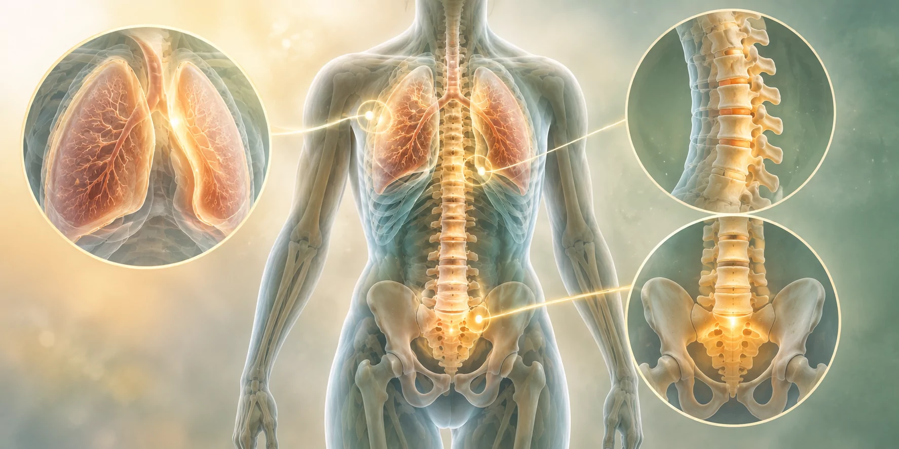 Considered together, these three experiences are particularly significant to me because they involved different structures and different manifestations:
- radiographically observed reduction or disappearance of spinal calcific changes;
- disappearance of disabling pain associated with L5–S1 spondylolysis and spondylolisthesis, despite spinal fixation surgery having previously been proposed;
- disappearance of longstanding pain associated with the aftermath of pleurisy.
I did not begin fasting with three separate instructions for my body.
I could not direct the process towards my spine, then towards the lumbar region, then towards the pleura.
The intervention was the same: I stopped supplying food from outside and allowed the organism to enter a profoundly different metabolic state.
The resulting changes appeared in different parts of the body and were neither selected nor consciously directed by me.
For this reason, the relationship between fasting, autolysis, autophagy and tissue remodelling remains deeply compelling to me. The metabolic change is systemic, while the processes occurring within individual tissues appear to follow priorities determined by the organism itself.
We may observe the consequences.
Understanding exactly how those priorities are established is the larger biological question.
Observation, Evidence and Mechanism
None of these experiences constitutes a controlled clinical trial, and I would not use them to claim that fasting will universally dissolve calcifications, resolve structural spinal disorders or eliminate the consequences of pleural inflammation.
At the same time, they cannot reasonably be reduced to vague subjective impressions.
In the first case, serial radiographs showed a change that several doctors independently recognised.
In the second, a disabling spinal condition for which surgery had been proposed was followed by years of pain-free, unrestricted movement.
In the third, a persistent symptom requiring regular analgesia disappeared and remained absent.
For me, these observations strongly suggest that fasting can be accompanied by processes of remodelling and recovery that deserve far more investigation than they currently receive.
Autophagy may explain part of what occurs. Changes in inflammatory signalling, tissue turnover, fluid balance, immune activity and other mechanisms may also contribute.
At present, the most rigorous position is to distinguish what was observed from the mechanism proposed to explain it.
The observations are real.
The biological explanation is still being explored.
Recycling, Adaptation and Renewal
Autophagy reveals a fundamental principle of living systems: survival and maintenance depend not only upon acquiring new material from outside, but also upon continually examining, dismantling and reusing what already exists within the cell.
During fasting, this internal economy becomes especially important. External nutrient availability decreases, cellular signalling changes and the organism must rely increasingly upon its own resources while continuing to preserve essential function.
Within that altered environment, some structures are retained, some are repaired and others may be dismantled so that their components can return to use.
The precision of these processes is remarkable.
At cellular level, damaged mitochondria can be selectively removed, protein aggregates can be recognised as targets and expendable material can be recycled while neighbouring structures remain intact. At the level of the whole organism, fasting likewise produces a hierarchy of resource use in which some reserves are readily mobilised while essential structures are defended for as long as possible.
How closely these two levels of selectivity are connected remains one of the most fascinating questions surrounding prolonged fasting.
The older Natural Hygiene concept of internal intelligence may use a different language from contemporary cell biology, yet both point towards the same striking characteristic of life: the organism constantly evaluates, adapts, preserves and renews itself according to changing conditions.
Fasting does not create this capacity. It profoundly changes the conditions under which it operates.
And perhaps this is one of the most important reasons to study autophagy: it allows us to see, at cellular level, something that fasting makes visible at the level of the whole organism — life sustaining itself through continuous adaptation and renewal.
